Summary
- Scientists at the Sudha Gopalakrishnan Brain Centre at the Indian Institute of Technology, Madras, have produced what they describe as the world’s most detailed three-dimensional atlas of the human brainstem at cellular resolution, offering an unprecedented view of one of the brain’s most vital yet poorly understood regions.
- Called Anchor, the atlas combines more than 500 tissue sections from foetal, childhood, and adult brains, allowing scientists to travel seamlessly from MRI scans of the whole brain to individual nerve cells.
- Built from high-resolution images of thin slices of post-mortem brain tissue, the approach makes detailed, cell-level mapping affordable, making it possible to chart the human brainstem at an unprecedented scale.
Scientists at the Sudha Gopalakrishnan Brain Centre at the Indian Institute of Technology, Madras, have produced what they describe as the world’s most detailed three-dimensional atlas of the human brainstem at cellular resolution, offering an unprecedented view of one of the brain’s most vital yet poorly understood regions. Called Anchor, the atlas combines more than 500 tissue sections from foetal, childhood, and adult brains, allowing scientists to travel seamlessly from MRI scans of the whole brain to individual nerve cells.
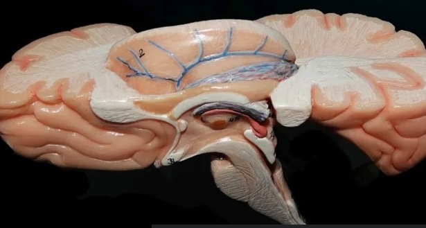
The brainstem occupies only a sliver of the brain, yet it keeps people alive by linking the brain to the spinal cord and controlling breathing, heartbeat, sleep, wakefulness, and movement. Damage to tiny clusters of cells within it can prove catastrophic, but the region’s densely packed architecture has long frustrated efforts to map it in detail. Anchor, built from high-resolution microscope images rather than costlier molecular techniques, identifies more than 200 clusters of brain cells and nerve pathways, using eight chemical markers to distinguish different cell types and producing one of the clearest pictures yet of this critical region.
Around 20 scientists spent 18 months at SGBC manually analysing more than 200 brain sections, combining MRI scans, microscopic anatomy, and 3D reconstruction into a single digital atlas. The centre now brings together more than 200 researchers, engineers, and technicians working with collaborators around the world. “Different atlases do different things,” said Mitra. MRI-based atlases capture the brain’s broad structure but not individual cells, while histological atlases map its architecture at cellular resolution. Anchor bridges these approaches, creating a reference library that could reveal how disease reshapes the brain cell-by-cell.
Scientists have mapped the brains of several animal species in remarkable detail, but the human brain remains comparatively under-charted because detailed studies of human brain tissue are scarce, Mohanasankar Sivaprakasam, who heads the SGBC, told the BBC. The centre now plans to image more than 100 whole human brains across different stages of life and neurological disorders, including Alzheimer’s disease and dementia. The atlas could also help explain how infections, including Covid-19, trigger long-term neurological damage and could help neurosurgeons navigate the brainstem more safely.
Part of Anchor’s appeal lies in its simplicity. Built from high-resolution images of thin slices of post-mortem brain tissue, the approach makes detailed, cell-level mapping affordable, making it possible to chart the human brainstem at an unprecedented scale. The achievement reflects a broader transformation in neuroscience, where progress increasingly depends as much on engineering and computation as on biology. As the centre continues its work, the atlas is expected to become a vital resource for researchers worldwide, advancing our understanding of the brain and its disorders.
We welcome your contributions! Submit your blogs, opinion pieces, press releases, news story pitches, and news features to opinion@minutemirror.com.pk and minutemirrormail@gmail.com